


MAP
Medial Anatomic Plate
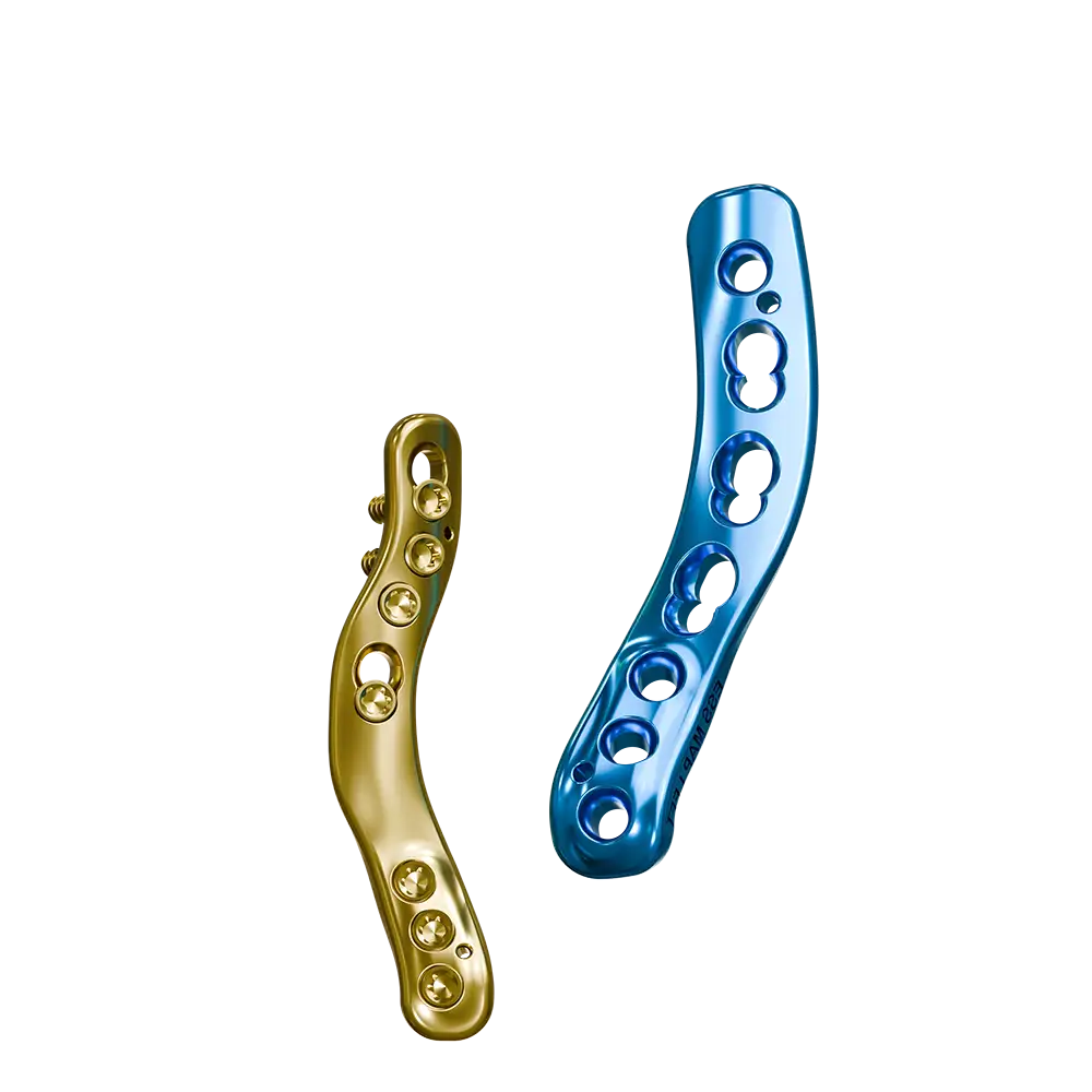
The Fusion Implants Medial Anatomic Plate (MAP) was developed through detailed CT-based mapping of the humeral topography of English Springer Spaniels (ESS) and French Bulldogs (FBD).
Designed using morphologic and biomechanical data, the MAP optimises screw placement and plate contouring to maximise bone purchase while minimising the risk of joint penetration, delivering reliable fixation across a range of fracture configurations.
The MAP is available in left and right configurations, with multiple plate lengths and two size variants derived from English Springer Spaniel and French Bulldog CT data, making it suitable for a range of small- to medium-sized dogs within this size category.
- Carefully pre-contoured distal end: curves cranially and over caudal aspect of medial epicondyle for maximum bone purchase.
- Sits more cranially than a straight plate, allowing for improved bone purchase with less risk of joint surface impingement.
- Midsection has a caudal “scoop” contour to minimise conflict with lateral plate (eg LEAP) screws when managing dicondylar (Y/T) fractures.
- Dense distal screw distribution typically allows placement of at least three screws distal to the fracture, even in very distal fractures, enhancing fixation security.
- Provides flexibility for locking fixation, dynamic compression, and lag screw placement, with variable angulation to reduce intra-articular or lateral screw conflict and support complex fracture management, including dicondylar (Y/T) fractures.

Demonstration Video
Training demonstration for both English Springer Spaniel MAP and French Bulldog MAP
MAP Range
Implants
Screws
Instruments
Courses
